Outline Diagram Of Animal Cell - Animal Cell Diagram Pencil Drawing Novocom Top / Animal cell cross section structure of a eukaryotic cell vector.. If so, you may need to memorize the animal cell, its organelles, and their functions. All animals, including you and i, are made of the same basic building block called the animal cell. An orange makes a great illustration of the three dimensional nature of cells. This was thought to be important to introduce the idea of a biological structure being adapted to its function to ensure. Animal cell cross section structure of a eukaryotic cell vector.
It is enclosed by two membranes in an envelope. The largest organelle within the cell. Let us look at animal cell parts and functions, using diagrams and illustrations. As observed in the labeled animal cell diagram, the cell membrane forms the confining factor of the cell, that is it envelopes the cell constituents together and gives the cell its shape, form, and existence. The diagram, like the one above, will include animal cells are eukaryotic in nature, possessing a nucleus and organelles that carry out the different functions the cell must do to thrive and reproduce.
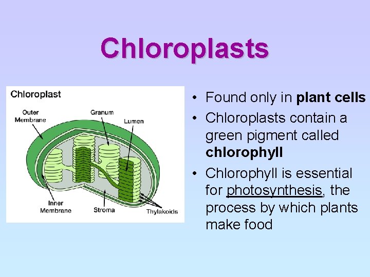
A Complete Guide To Draw Biology Diagrams In Easy Steps Biology Diagrams Plant Cell Plant Cell Diagram from i.pinimg.com A comparison of plant and animal cells using labelled diagrams and descriptive explanations. The most important structures of plant and animal cells are shown in the diagrams below, which provide a clear illustration of how much these cells have in common. Most cells are very small; The diagram, like the one above, will include animal cells are eukaryotic in nature, possessing a nucleus and organelles that carry out the different functions the cell must do to thrive and reproduce. A system of flattened membranes called cisternae (mainpoint: Cut the orange in half. How do you tell eukaryotic cells from prokaryotic cells? I spelt it wrong in the diagram, sorry).
An orange makes a great illustration of the three dimensional nature of cells.
Cut the orange in half. Make a checklist of all of the organelles, and check them off as you draw them to ensure you have not left anything out of your diagram. Each organelle has a different purpose inside the cell. The diagram, like the one above, will include animal cells are eukaryotic in nature, possessing a nucleus and organelles that carry out the different functions the cell must do to thrive and reproduce. In truth, there are still features of plant and animal cells we're only lately. During animal cell division, the centrioles replicate (make new copies) and the centrosome divides. Cells are the smallest units of life. Smooth endoplasmic reticulum, mitochondria, golgi bodies, lysosomes. There are three microtubules in each group. I spelt it wrong in the diagram, sorry). Here is a summary of their structure and function. It helps in carrying out the functions such as respiration, nutrition, digestion, excretion etc. Both plant and animal cells are surrounded by a cell membrane composed of lipids and proteins.
Start studying animal cell diagram. Animal cells are generally smaller than plant cells and lack a cell wall and chloroplasts; Diagram showing anatomy of animal cell. Generalized cell is used for structure. Cut the orange in half.
Animal Cell Stock Illustrations 9 514 Animal Cell Stock Illustrations Vectors Clipart Dreamstime from thumbs.dreamstime.com You know, animal cell structure contains only 11 parts out of the 13 parts you saw in the plant cell diagram, because chloroplast and cell wall are available only in a plant cell. Diagram showing anatomy of animal cell. Prokaryotic cells (above) are much simpler in structure than eukaryotic cells. Animal cell structure animal cells are typical of the eukaryotic cell, enclosed by a plasma membrane and containing a membranebound nucleus and organelles. But at the same time it is interpretive. All the living organisms are made up of cells and it is the smallest unit of life. There are three microtubules in each group. Each centriole is a ring of nine groups of fused microtubules.
The role and function of the plasma membrane;
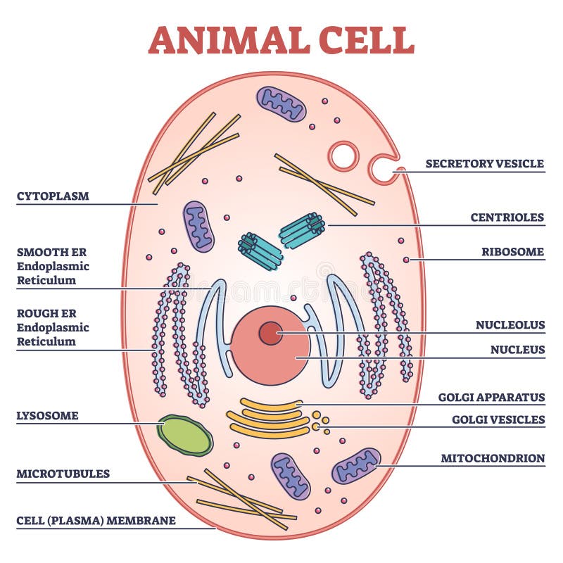
Animal cell with labeled anatomic structure parts diagram outline concept. This is where the digestion of cell. The largest organelle within the cell. The animal cell diagram on the free worksheet will teach students to identify the function of the major parts of the animal cell. Both plant and animal cells are surrounded by a cell membrane composed of lipids and proteins. The cell (from latin cella, meaning small room) is the basic structural, functional, and biological unit of all known organisms. Animal, plant, protist, and fungus cells. Later we look at the hierarchical organisation of cells into tissues then into organs then into systems and then into an organism. Lets us discuss the animal cell, types of an animal cell, animal cell diagram, its structure. Human worm icon, outline style. If so, you may need to memorize the animal cell, its organelles, and their functions. Cytoplasm, ribosomes, rough endoplasmic reticulum; All animals, including you and i, are made of the same basic building block called the animal cell.
An animal cell diagram is a great way to learn and understand the many functions of an animal cell. During animal cell division, the centrioles replicate (make new copies) and the centrosome divides. Make a checklist of all of the organelles, and check them off as you draw them to ensure you have not left anything out of your diagram. Each cell can be thought of as a large factory. So it is called as the structural and.
Cell Structure Organisation Chapter Outline A Identify Cell from slidetodoc.com All organisms are made up of cells (or in some cases, a single cell). But at the same time it is interpretive. There are three microtubules in each group. The result is two centrosomes, each with its own pair of centrioles. All animals, including you and i, are made of the same basic building block called the animal cell. If so, you may need to memorize the animal cell, its organelles, and their functions. That's the major difference between plant and animal cells under microscope. So it is called as the structural and.
The animal cell diagram on the free worksheet will teach students to identify the function of the major parts of the animal cell.
An animal cell diagram is a great way to learn and understand the many functions of an animal cell. Animal cells are the basic unit of life in organisms of the kingdom animalia. The nerves and muscles are made up of specialized cells that plant cells. I spelt it wrong in the diagram, sorry). The plant cell wall gives the cell a lot of strength and prevents it from bursting under high pressure as it is made up of cellulose arranged in groups called microfibrils. Cytoplasm, ribosomes, rough endoplasmic reticulum; The animal cell diagram on the free worksheet will teach students to identify the function of the major parts of the animal cell. Both plant and animal cells are surrounded by a cell membrane composed of lipids and proteins. Since animal cells lack a rigid cell wall it allows them to develop a great diversity of cell types, tissues, and organs. Human worm icon, outline style. A system of flattened membranes called cisternae (mainpoint: 5th grade science and biology. To help you do this, i've created a printable animal cell diagram.
Berbagi :
Posting Komentar
untuk "Outline Diagram Of Animal Cell - Animal Cell Diagram Pencil Drawing Novocom Top / Animal cell cross section structure of a eukaryotic cell vector."


Posting Komentar untuk "Outline Diagram Of Animal Cell - Animal Cell Diagram Pencil Drawing Novocom Top / Animal cell cross section structure of a eukaryotic cell vector."